
Application & technical notes
Explore Inscoper’s microscopy application and technical notes — practical insights, imaging techniques, and real-world fluorescence microscopy use cases. Discover how our expertise in device control and image acquisition software can help you achieve sharper, faster results.
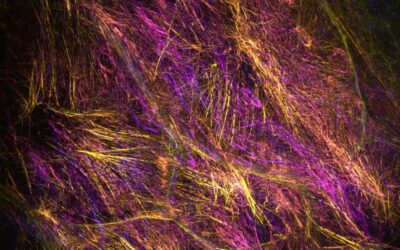
Overcome a paradoxical situation in a complex biological context using dynamic Random Illumination Microscopy
Contraction vs expansion of actomyosin networks in deep epithelia of drosophila How to overcome a paradoxical situation in a complex biological context using dynamic Random Illumination Microscopy Introduction: The challenge of dynamic tissue high resolution imaging...
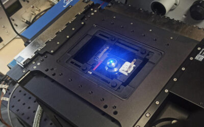
Higher XY Speed for Live Cell Imaging with ZABER Nucleus microscope
Summary Nucleus motorized microscopes from Zaber form a compact benchtop microscope platform that offers a flexible configuration to adapt the user needs, and includes fast and precise motorized XY and Z-focus movement devices. Nucleus microscopes are fully controlled...
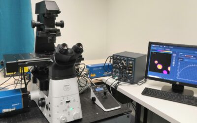
FD-FLIM imaging with pco.flim camera system
Fluorescence phenomenon in microscopy can be characterized by its intensity, but also by the time between the excitation of a fluorescent molecule and the emission of the corresponding photon. This measure of time is a microscopy technique called FLIM, for...
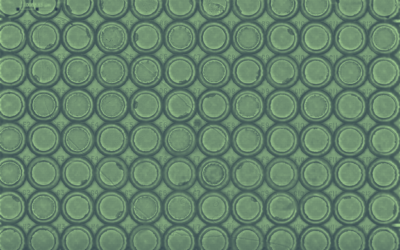
Continuous Motion Imaging for Very High-Speed Microscopy
Microscopic imaging of a large number of biological samples is needed for a wide spectrum of applications in life sciences, including High Content Screening (HCS) protocols. However, conventional microscopy is limited by some technical limitations (movement speed of...
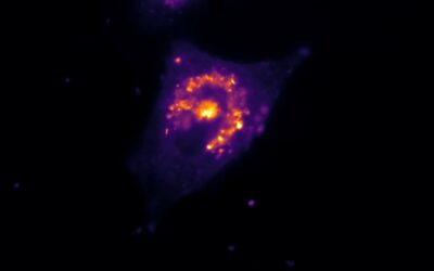
Characterization of organelle dynamics in living cells by combining Oxxius lasers and Inscoper scanFRAP
Fluorescence Recovery After Photobleaching (FRAP) is nowadays considered as the mode widely used method to monitor protein and organelle dynamics. This application note introduces the use of the Inscoper scanFRAP solution to explore the cinetic of endoplasmic...
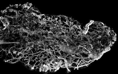
Imaging cleared organ with an Ultramacroscope using Inscoper Imaging Solution
Light-sheet fluorescence microscopy is an illumination technique well-suited for volumetric imaging that can be used for a wide spectrum of biological applications. Here, we present an opportunity for researchers to upgrade their macroscope to an “ultramacroscope”, a...
Monitoring of kinase activation using FRET biosensor on living cells with Inscoper fastFLIM
Inscoper fastFLIM applications The Inscoper fastFLIM, a camera-based time-domain fluorescence lifetime imaging microscopy (FLIM) system, is the fastest lifetime imaging technique designed for live cell experiments. This technology can be used : to follow molecular...
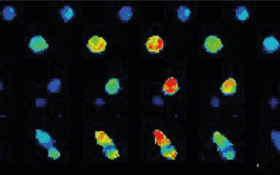
Live calcium imaging to monitor T cells activation using Inscoper liveRATIO
When stimulated by the environment, many cell types use calcium signals for intracellular processing of information and induction of appropriate biological responses through activation of specific gene expression programs. For instance, calcium transients are...
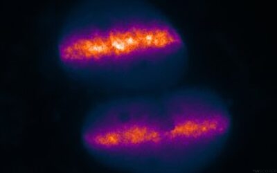
Microirradiation using Inscoper scanFRAP for real-time DNA repair monitoring
Maintenance of the genome integrity is directly dependent on the spatiotemporal recruitment and regulation of the repair proteins at DNA damage sites. The alteration of this complex biological pathway could induce mutations or premature cell death. The real-time...

Confocal microscopy with Inscoper Solution using Confocal.nl technology
Live-cell microscopy is nowadays widely used to image and better understand the fundamental nature of cellular function and organization. The development of diversified microscopy approaches, such as confocal microscopy, allows to image a large spectrum of cellular...
Laser-induced DNA damage within a 4D/5D acquisition sequence
INSCOPER markets a laser illumination controller that allows photomanipulation experiments to be performed on a video microscope within a multidimensional acquisition sequence. This 4' video shows an image acquisition with 3 sequences: before, during and after laser...
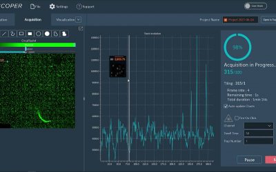
Image Acquisition & Tiling
The Tiling feature included in the INSCOPER Microscopy Imaging Solution is used to image an entire slide (or well) or a large region of interest in the sample. Several images are acquired one after one in a row, called “tiles”, then “stitched” afterward to constitute...
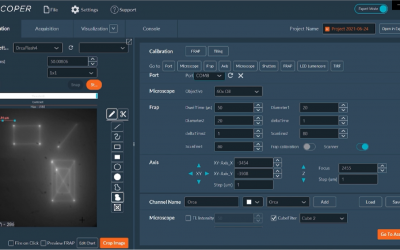
FRAP Calibration
For the optimal use of the Photomanipulation Dimension of the INSCOPER Imaging Software, it is recommended to calibrate the laser in order to match up the position of the scanner optical head with the camera screen size. The microscope system is equipped with FRAP...
Image acquisition software is the microscope’s brain
Fluorescence microscopy has been a genuinely disruptive innovation for examining live specimens. It has enabled the invention of numerous imaging techniques and methods for observing the behaviour of biological objects of interest in a way that mirrors their spatial...

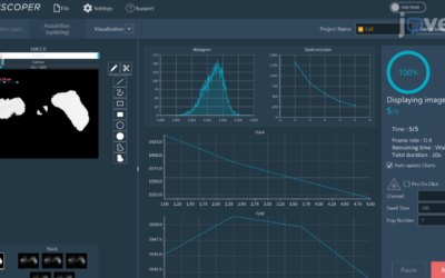
How to set time lapse
The procedure is as follows: Adjust the clarity of the image. Select a channel or add a new one. Determine the number of time points. Set an interval between each time point – >Total time will be calculated automatically. If other settings are not needed, disable...