the easy-to-use flim solution for liVe cell imaging
Inscoper fastFLIM is a turnkey solution with real-time calculation of the lifetime image, compatible with all brands of microscopes. It can be used with any type of illumination in camera detection such as wide field, spinning disk, TIRF, SPIM.
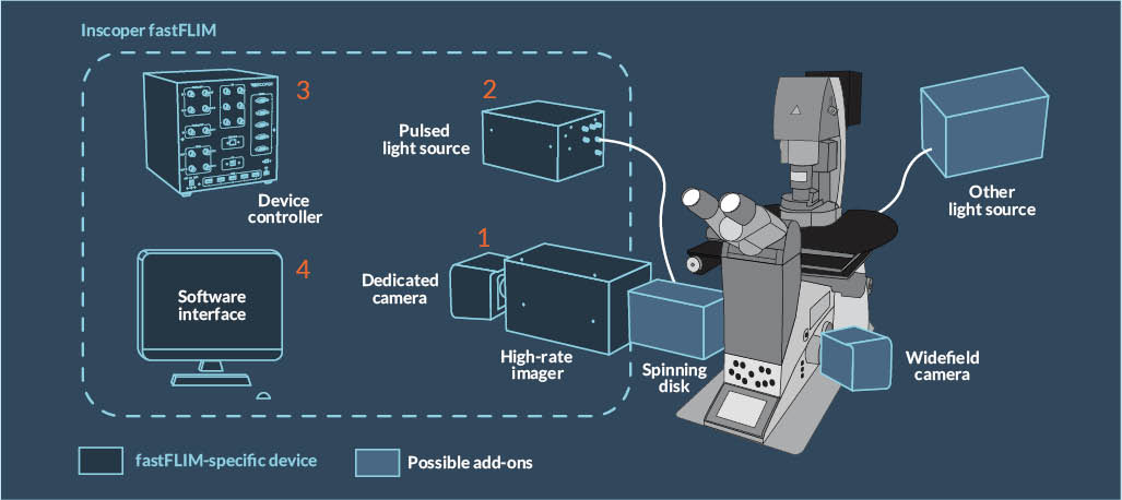
The fastFLIM solution consists of:
(1) an optical module that connects to the camera port of a conventional microscope body, composed of a high-rate imager (to generate time gates) and a scientific camera,
(2) a pulsed white laser with a wavelength selection system,
(3) a device controller for triggering and synchronizing the microscope signals,
(4) a complete image acquisition software.
the first time-domain flim for camera microscopes
The Inscoper fastFLIM solution is perfectly suited for live cell imaging. It can be used for a large panel of applications, including lifetime monitoring, FRET measurement (protein-protein interactions), biosensor analyzes, or control of the environment in the sample (pH, viscosity, etc.).
With its simplicity, fastFLIM democratizes the use of FLIM as a complementary measurement to quantitative fluorescence imaging. It is fully integrated into the Inscoper Imaging Solution to offer a robust and user-friendly product that can be combined with any other fluorescence imaging technique and third-party equipment.
USER CASE @ RENNES, FRANCE
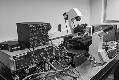
The microscopy system combines the fastFLIM solution in addition to the Inscoper scanFRAP, which is a dedicated solution for laser photomanipulation. It equips a microscopy core facility, where users are regular biologists with various levels of practice in microscopy.
perfectly suitable for multidimensional acquisitions
Seamless and complete integration of the microscope
The third party devices that equip the microscope are fully controllable: cameras from leading manufacturers such as Andor, Hamamatsu, PCO and Photometrics, microfluidic devices (pumps, temperature…), light sources and filter wheels, XYZ stages, etc.
real-time imaging for low photo-toxicity
SUPPORT SERVICE
We can frequently optimize the system rapidly using remote tools but we are available to work on your site when needed.
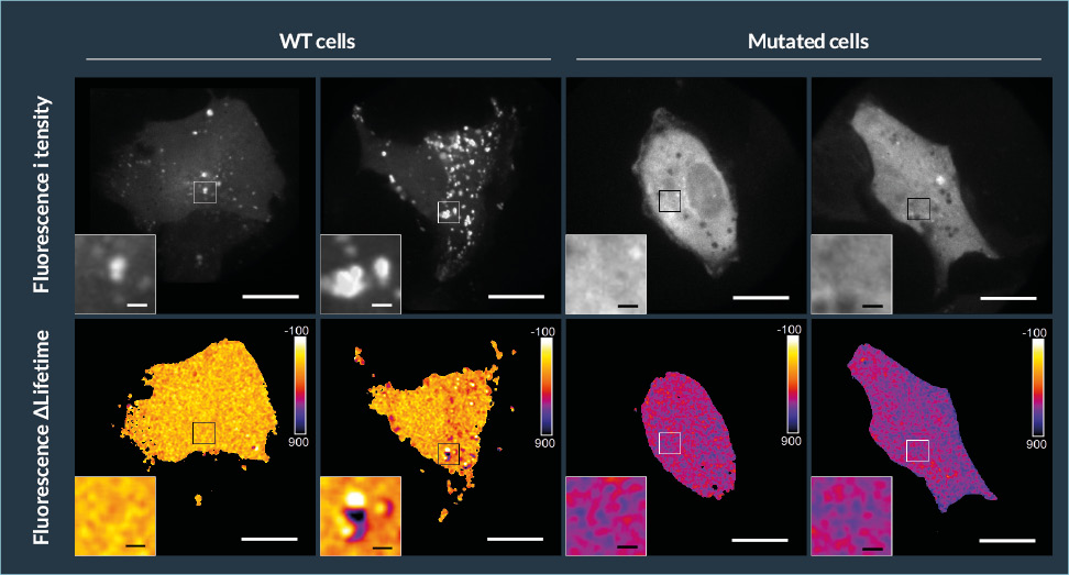
Biosensor imaging with FRET/FLIM using the Inscoper fastFLIM
Representative fluorescence intensity and ΔLifetime images of WT and mutated U2OS cells, co-expressing biosensor and analyzed by FRET/FLIM. Squares on the images illustrates the location of the zoomed images. Pseudocolor scale: pixel-by-pixel ΔLifetime. Scale bars: 40 µm (large images) and 5µm (enlarged images).
Courtesy of Dr. Elif Begüm Gökerküçük, IGDR, Rennes, France

innovative technology for live & automated FLIM imaging
fastFLIM renews the way to do FLIM imaging on a camera microscope. The FLIM measurement is done in time domain, with a picosecond pulsed laser and a time gate generator. It has the advantages of doing FLIM with a camera detection, namely the low photo-toxicity and the high acquisition speed, but without the need to use a reference sample or a fitting calculation to perform the measurement.
Temporal gates of 2 ns in a time window of 10 ns are sequentially generated to obtain a stack of 5 time-gated images. These images are then used to calculate the pixel-by-pixel mean fluorescence Δlifetime in real time, according to the following equation: τ = ΣΔti x Ii /ΣIi, where Δti corresponds to the delay time of the ith gate while I indicates the pixel-by-pixel time-gated intensity image.
This method ensures rapid FLIM measurements: no fitting or binning steps are required, and lifetime is calculated in live, with minimal photon budget.
Easily accessible for regular biology users
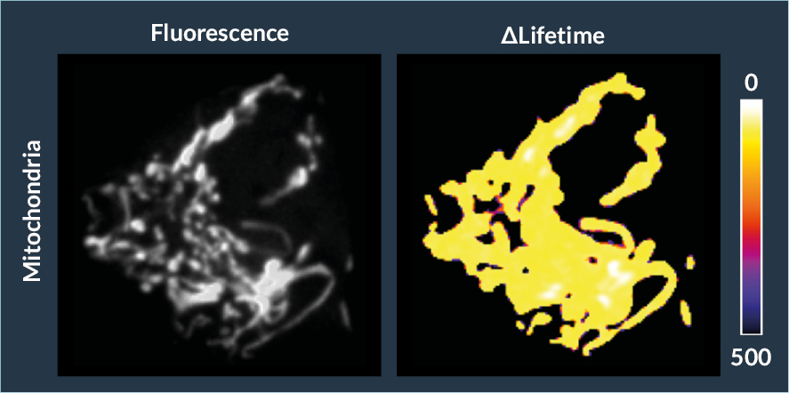
Mitochondria monitoring with FLIM/FRET using biosensors
Forster’s Resonance Energy Transfer by Fluorescence Lifetime Imaging Microscopy analyses on MCF7 cells expressing a fluorescent biomarker. Fluorescent-labeled mitochondria are image according their fluorescence intensity (left) and ΔLifetime (right)
Courtesy of Dr. Giulia Bertolin, IGDR, Rennes, France
To make FLIM available to cell biologists, we needed a simple solution that could be automatically combined with other microscope imaging modalities.
The fastFLIM solution addresses the growing need for rapid lifetime imaging to solve many of the challenges brought by the scientific teams at our institute.
With this system our users are completely autonomous, which allows for a better workflow on our microscopy imaging facility.
No preparation required
There is no need for a reference sample, users just have to position their sample and launch the acquisition of images.
No complicated calculation
Users do not have to do the math, the result is direct as there is no fitting calculation to proceed.
Fast
Five intensity images are enough to calculate one FLIM image, allowing an average frame rate of 2 to 10 images FLIM images per second (depending on the exposure time).
LIVE
FLIM images are immediately displayed and processed in real time.
Reproducible
The measurement is perfectly stable over time, whatever the duration and repetition of the acquisitions.
Compatible
fastFLIM includes the Inscoper Imaging Solution that can make this convenient FLIM technique a stand-alone microscopy solution or an additional imaging modality.
FLIM-FRET experiments using Inscoper fastFLIM
Real-Time Monitoring of Aurora kinase A Activation using Conformational FRET Biosensors in Live Cells
JoVE Journal Biology, DOI : 10.3791/61611-v
related scientific publications
