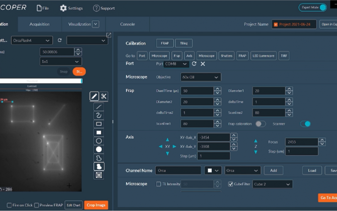
Image Acquisition & Tiling
The Tiling feature included in the INSCOPER Microscopy Imaging Solution is used to image an entire slide (or well) or a large region of interest in the sample. Several images are acquired one after one in a row, called “tiles”, then “stitched” afterward to constitute the whole sample.
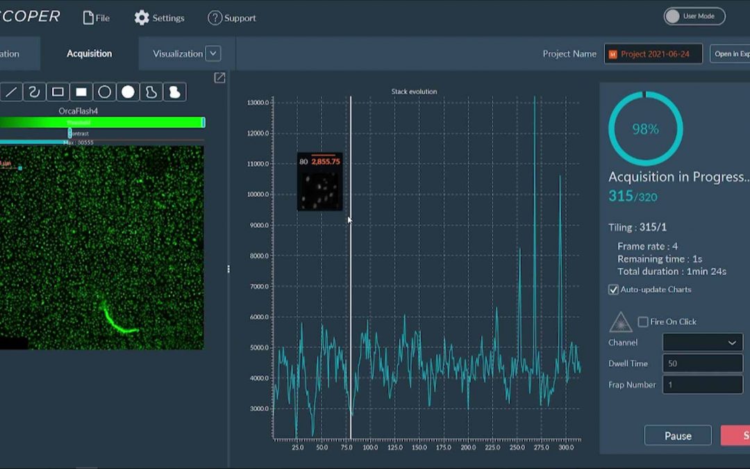
This tutorial shows how to set up the acquisition sequence and proceed to the tiling of U2OS cells transfected with ALC1-GFP and labeled with Hoechst at 37°C. The specimen is placed in an 8-chambres glass holder.
The steps are as follows:
- Select the illumination channel.
- Switch the “Positions” dimension to “Tiling”
- Define the parameters for tiling imaging:
- Select 2 key points, on diagonal;
- Set the overlap and define the read mode.
- Set the channel in the Multi-Channels dimension.
- Select the data processing algorithm.
- Choose the storage location for the dataset.
- Start the acquisition & confirm.
- Monitor the progress of the acquisition.
- Visualize your data.