
Dynamic Super-Resolution
No more trade-offs for live cell & thick tissue imaging
For decades, researchers have been forced to choose between resolution, speed, and sample viability.
liveDRIM™ shatters these boundaries, delivering super-resolution to live-cell imaging
without the typical trade-offs that limit live imaging.
See Sharper
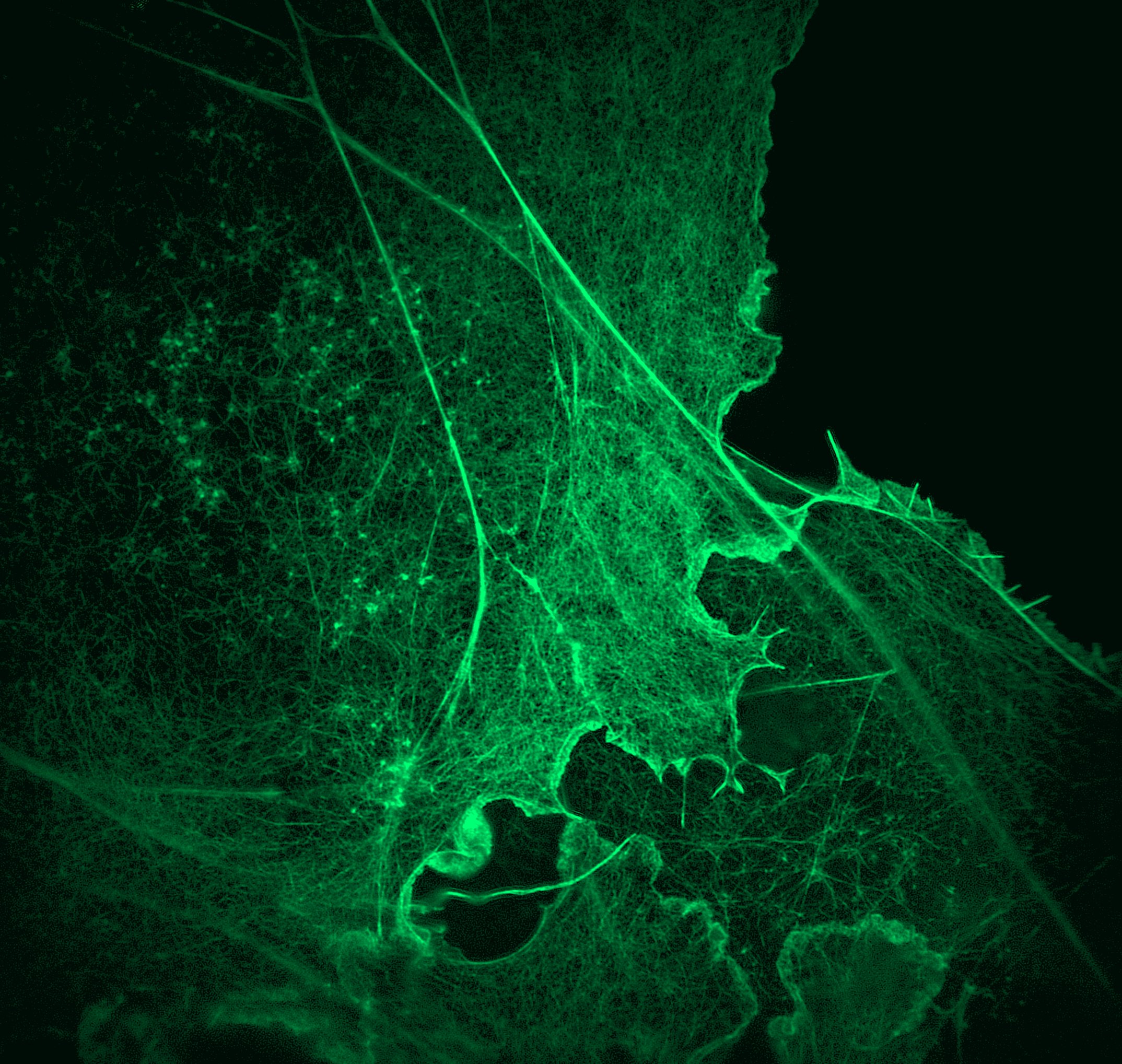
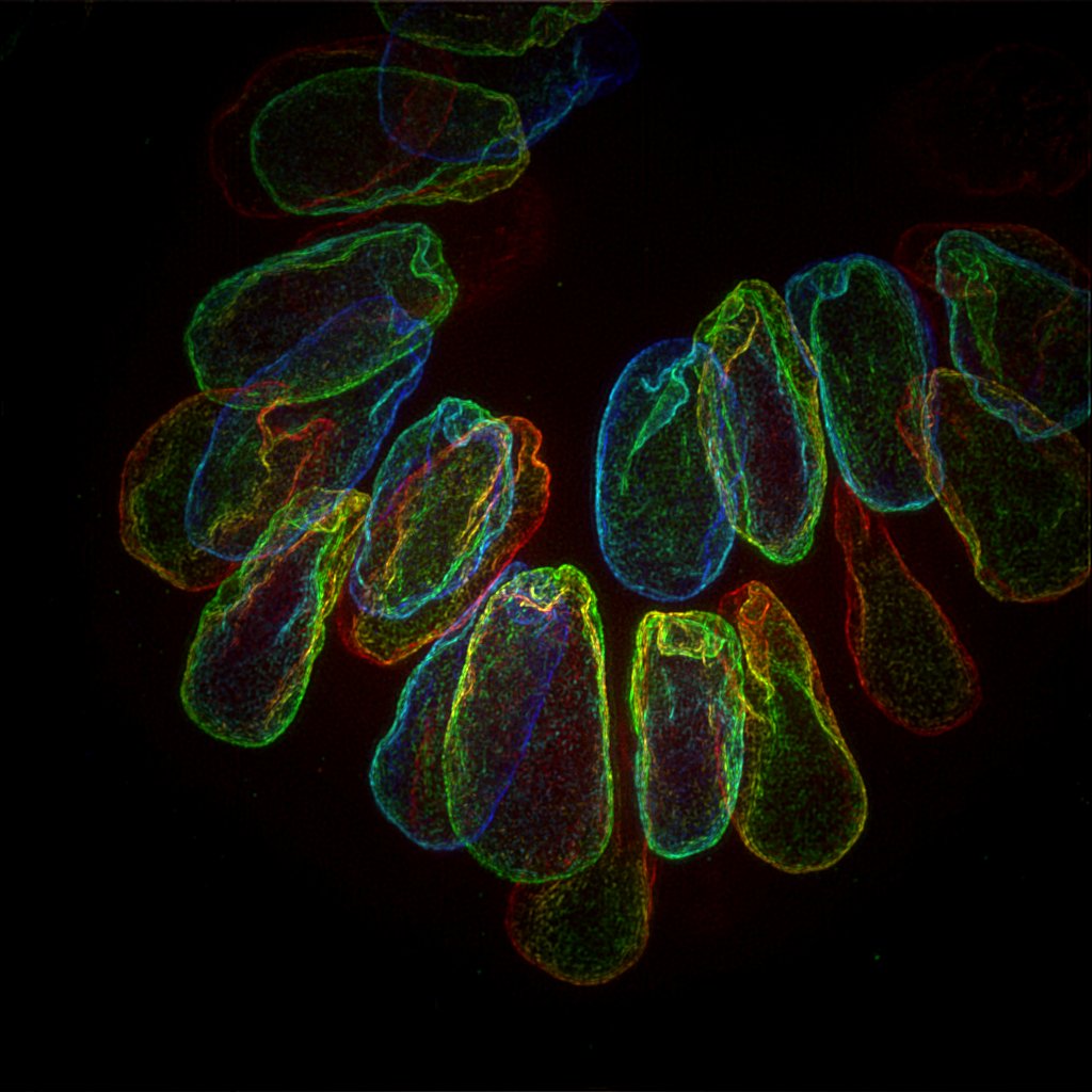
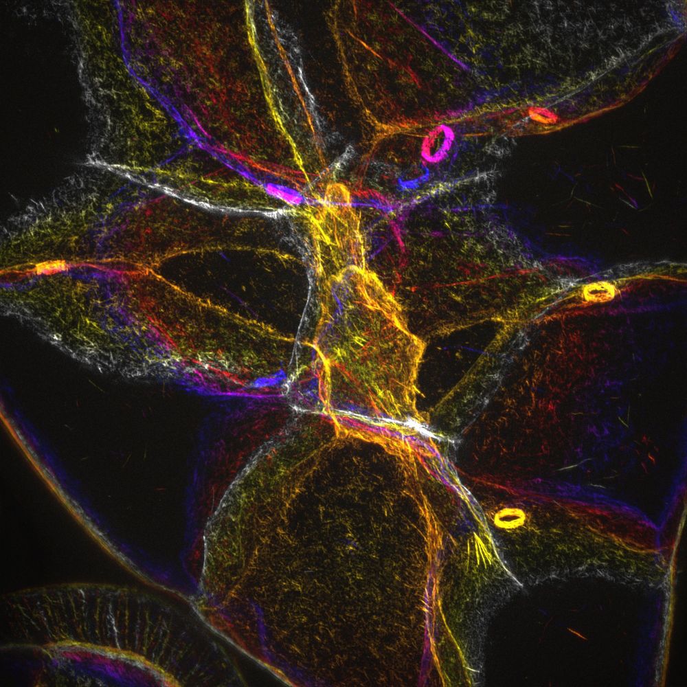
Achieve 90-130 nm XY resolution in multiple colors. Reveal structures previously hidden by the diffraction limit.
Explore Deeper
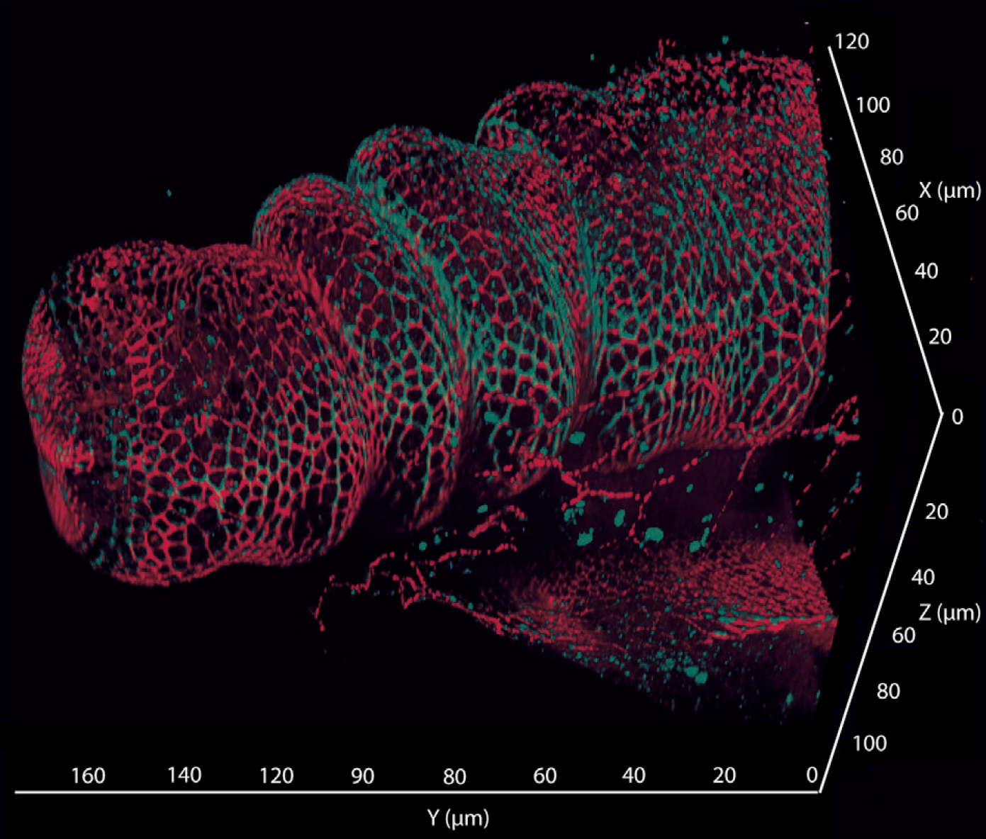
Image up to 100 µm in depth with artifact-free clarity. Perfect for organoids, embryos, and thick tissue sections.
Capture Faster
With speeds up to 12.5 frames per second, you can track dynamic biological events as they happen, not after they’ve passed.
Observe Long-term
Engineered for low photo-toxicity, liveDRIM protects your samples, enabling long-term time-lapse studies without compromising cell health.
Designed for demanding researchers
liveDRIM™ isn’t just a new imaging mode; it’s a seamless addition to your existing microscopy workflow. We’ve removed the technical hurdles that typically come with super-resolution.
- No special sample prep: Use your standard fluorescent markers and sample holders (slides, multi-well plates). No specialized buffers required.
- Unmatched versatility: Fully compatible with all regular acquisition features, including multi-dimension (3D + Time), multi-color imaging.
- Full Field of View: Our camera-based technology ensures you don’t sacrifice area for detail.
- Instant Feedback: Use the “Live” imaging mode to see liveDRIM results immediately, allowing you to optimize your experiment before you even hit ‘Acquire’.

Watch as we compare standard Widefield imaging against liveDRIM to show the dramatic leap in clarity and depth.
Uncompromising technology: How we compare
How does LiveDRIM™ stack up against the imaging modalities you know? We’ve engineered a “sweet spot” that combines the depth of Confocal with the resolution of SIM and the gentleness of Light-Sheet.
| Microscopy Technique | XY resolution (nm) | Acquisition speed | Phototoxicity | Optical Sectioning | Depth Imaging Robustness* |
|---|---|---|---|---|---|
| Two-Photon | 400 | - | Low | Good | ++ |
| Widefield | 200 | ++ | Low | Poor | - |
| Confocal laser scanning | 200 | - | High | Good | ++ |
| Confocal spinning disk | 200 | ++ | Moderate | Good | + |
| Light-Sheet | 200 | ++ | Low | Good | + |
| SIM | 100 | + | Moderate | Moderate | - |
| liveDRIM | 100 | + | Low | Good | ++ |
| STED | 20 | - | High | Moderate | + |
| PALM/STORM | 10 | - | High | Poor | - |
* imaging quality on thick scattering samples
Case Study: Solving the “Actomyosin Paradox”
How liveDRIM revealed the hidden dynamics of Drosophila morphogenesis
In the study published in Nature Communications, researchers, Wang et al. were able to observe deep within the curved follicular epithelium of Drosophila to uncover a completely new mode of actomyosin behavior by using liveDRIM.
The liveDRIM difference: Deep, Dynamic, Delicate
Imaging the basal side of a curved tissue behind multiple scattering layers typically causes standard super-resolution (SIM) to fail. liveDRIM provided the only modality capable of answering all researchers’expectations simultaneously:
- Superior depth: Unlike traditional SIM, our Random Illumination is robust against optical aberrations, maintaining 100 nm resolution even through scattering tissue.
- Nanoscale clarity: Revealed that these specific stress fibers were non-linear and fragmented—explaining why they expanded instead of contracted.
- Temporal fidelity: Captured fast cytoskeletal pulses (every 30-120s) and focal adhesion assembly in real-time.

Ready to transform your imaging?
Every imaging challenge is unique. Whether you are working with organoids, embryos, or complex tissue sections, our team is here to help you determine when liveDRIM™ is the right fit for your research goals.
Schedule a technical consultation to discuss:
- Sample compatibility and preparation.
- Integration with your current microscope setup.
- A personalized remote demo using your own parameters.
The science behind the solution:
A strong partnership
liveDRIM™ is a collaborative breakthrough in fluorescence microscopy and automation:
- Manufactured by Gataca-Systems: Precision-engineered optical modules for seamless hardware integration.
- Patented technology by Rimeo: Based on the published Random Illumination Microscopy method. (Mangeat et al., Cell Reports Methods, 2021).
- Powered by Inscoper: The intelligent control system orchestrating high-speed image acquisition and synchronization between devices ( liveDRIM, cameras, lasers and any peripherals).
