
What can super-resolution microscopy bring to researchers working with live cells, thick tissues and other complex biological samples?
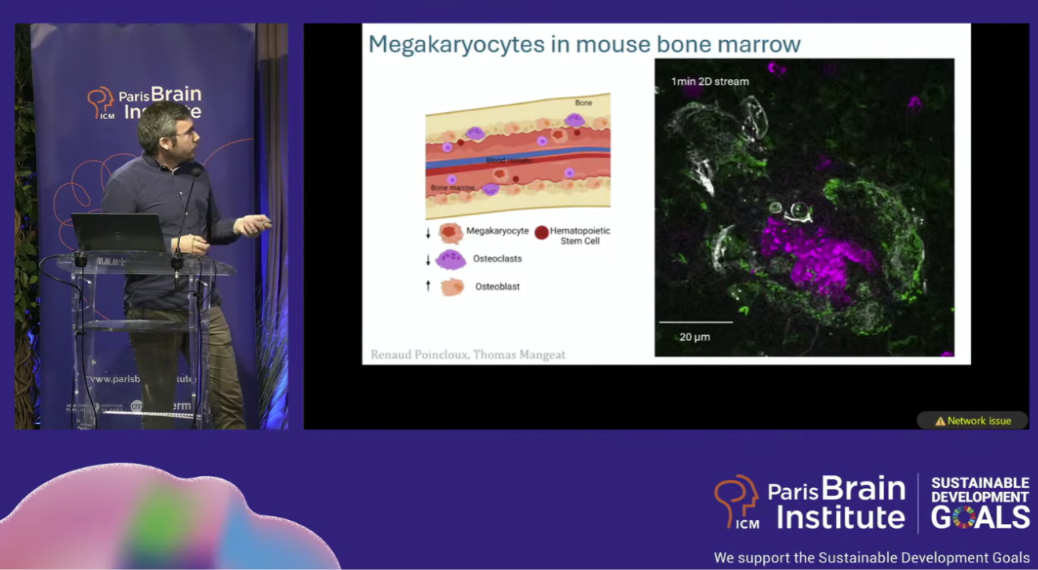
At the 5th edition of TED ImaBio 2026, Simon Labouesse from Rimeo presented liveDRIM (live Dynamic Random Illumination Microscopy), our super-resolution imaging solution designed for live and complex biological samples.
His presentation shows how liveDRIM combines super-resolution, fast acquisition, low phototoxicity and imaging at depth, while providing researchers with immediate visual feedback during their experiments.
Watch the 5th edition of TED ImaBio. Jump to 1:25:00 for Simon’s talk (note that the presentation is in French).
liveDRIM for live and deep super-resolution imaging : Key take aways from the conference
- No grid artifacts in deep tissue: Uses Random Illumination Microscopy (RIM) and speckle covariance to remain immune to optical aberrations, reaching up to 130 µm depth.
- Instant visual feedback: Real-time processing displays super-resolved images on-screen instantly, making it effortless to find your ROI and adjust focus live.
- Nanoscale precision at high speed: ~90 nm XY resolution at up to 12.5 Hz (4–5 ms/plane), far faster than point-scanning confocals.
- Gentle on live samples: Ultra-low phototoxicity ideal for sensitive time-lapses (e.g., tracking 3D FtsZ ring closure or cell migration).
- Zero-hassle calibration: “Releasing the steering wheel” removes rigid illumination constraints and daily setup friction for core facility users.
- Expansion microscopy compatible: Pairs seamlessly with expansion protocols to resolve dense nanostructures (like 250 nm centrioles).
Unlike Structured Illumination Microscopy (SIM), which relies on precisely controlled illumination patterns, the Random Illumination Microscopy approach uses a sequence of random patterns and reconstructs the super-resolved image statistically from the resulting camera images.
This approach can achieve a resolution gain comparable to SIM while being more robust to optical aberrations that can distort illumination patterns when imaging deeper into complex samples.
For researchers, liveDRIM translates these properties into a practical imaging solution combining high spatial resolution with fast, low-phototoxicity imaging, including in conditions where conventional structured illumination becomes more challenging.
Why choose liveDRIM for live and deep super-resolution imaging?
As Simon explains during the presentation, one of the fundamental differences between SIM and the technology behind liveDRIM lies in how the illumination pattern is handled.
In conventional SIM, reconstruction relies on knowing the structured illumination pattern applied to the sample. As imaging depth increases, optical aberrations and scattering can distort this pattern. The reconstruction algorithm, however, still expects the original illumination, which can introduce artifacts.
The technology behind liveDRIM takes a different approach: the exact illumination pattern does not need to be known. Instead, reconstruction relies on the statistical properties of the random illumination.
This makes the method more tolerant to distortions of the illumination pattern and particularly relevant for super-resolution imaging in thick or complex biological samples.
From bacteria to deep live-tissue imaging
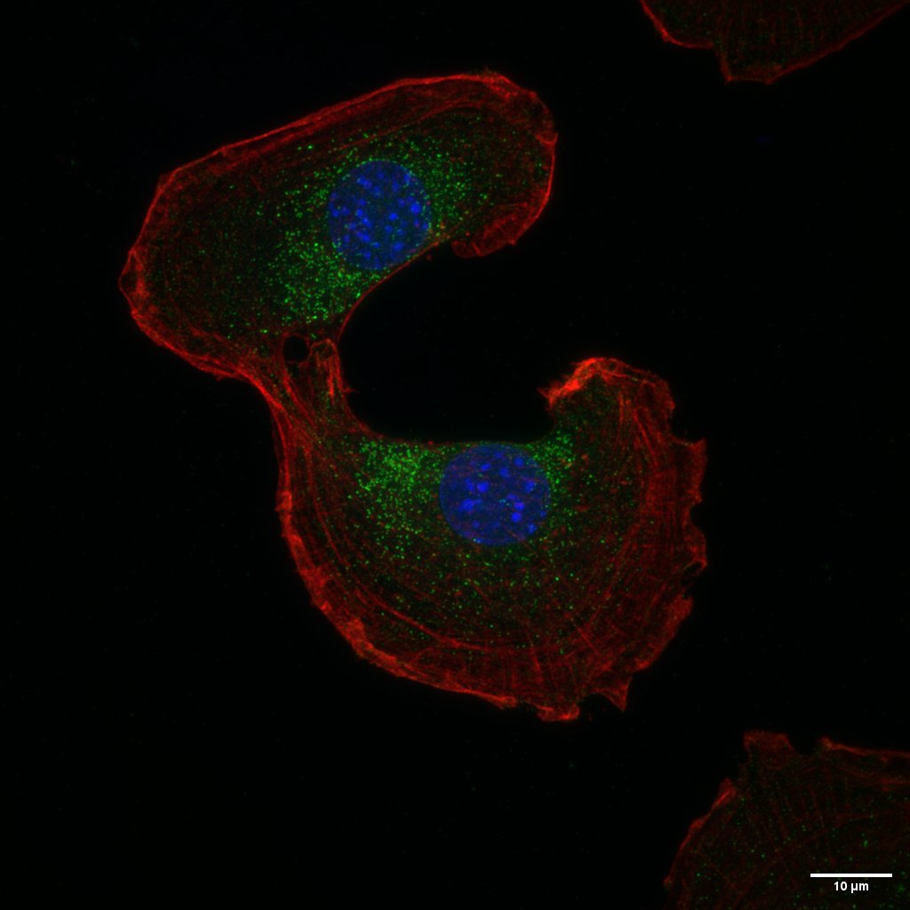
The TED ImaBio presentation illustrates liveDRIM across very different biological samples and experimental requirements.
Simon presents examples including bacterial imaging, expansion microscopy, multicolor imaging and fast imaging of migrating cells in Drosophila tissue. The presentation also demonstrates super-resolution reconstruction at different imaging depths in complex samples.
These examples illustrate why combining resolution with acquisition speed, low phototoxicity and robustness at depth matters for biological imaging: the objective is not simply to produce a sharper image, but to observe spatial detail while preserving access to the dynamics of the biological process.