
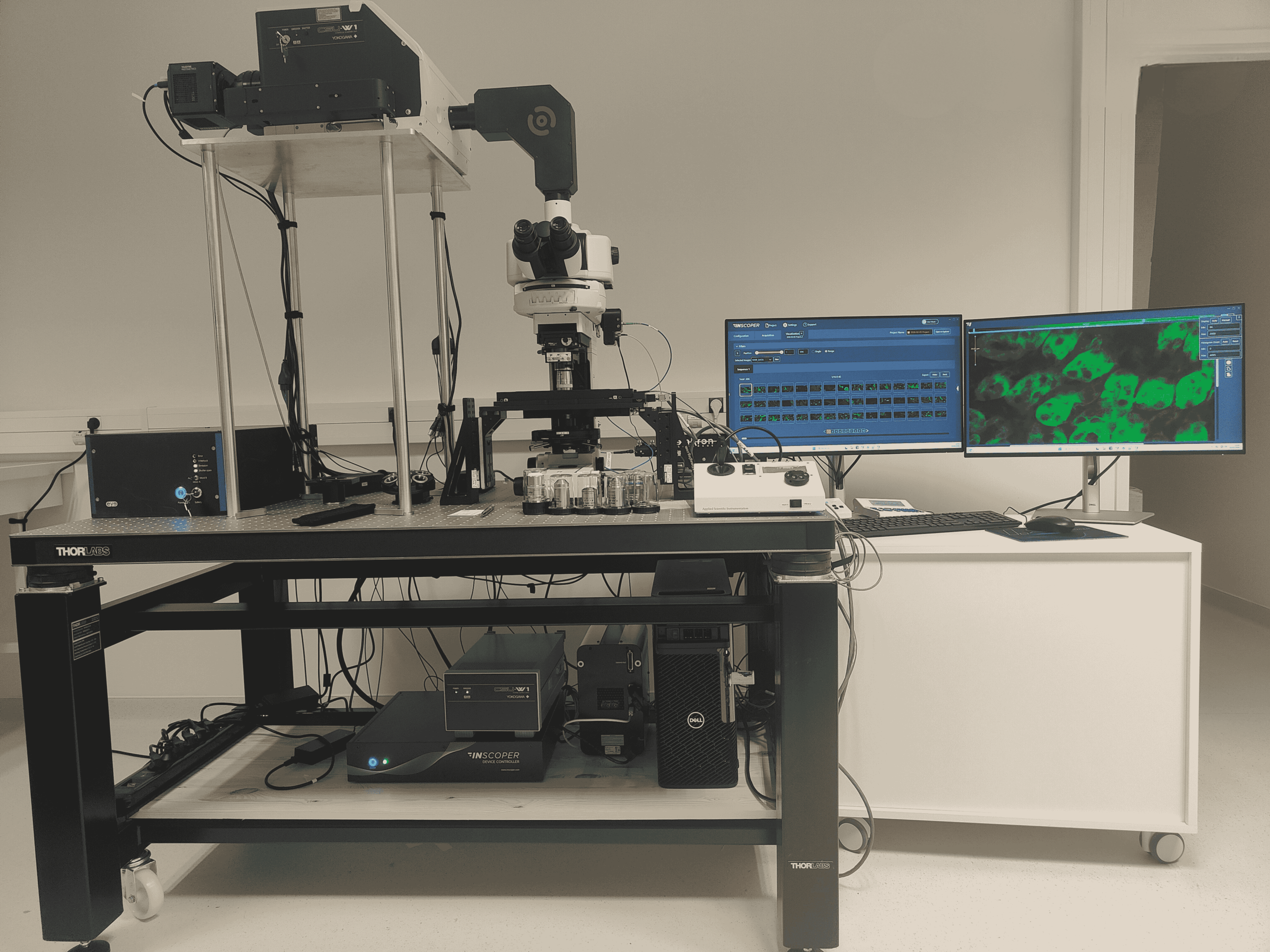

System description & applications
This system is used for intravital microscopy in mice to study the brain and heart in vivo.
Image acquisition can be synchronized with the animal’s respiratory and cardiac cycles to minimize motion artifacts and reduce blurring, ensuring high-quality data while monitoring physiological parameters in real time.
This setup enables precise observation of physiological processes in living tissues. It is used for intravital imaging of moving organs, particularly the heart, to investigate immune cells involved in chronic cardiac diseases.
Additional applications include imaging of live and fixed organoids and spheroids, as well as the development of ratiometric imaging approaches enabled by the system’s dual-camera configuration.

microscope device list
-
-
Microscope: Upright Nikon microscope
-
Stage: PRIOR motorized Z-stage
-
Camera: Two Photometrics Kinetix sCMOS cameras
-
Spinning disk: Yokogawa W1 T2
-
Illumination: PE-4000 fluorescence light source
-
Laser system: CAIRN laser bench (405 / 488 / 561 / 640 / 690 / 730 nm)
-
Imaging module: SPARQ module (Bliq Photonics)
-